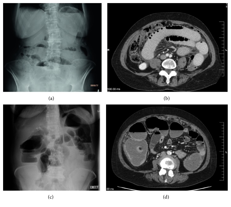
Figure 1.
Radiographic images showing malignant bowel obstruction. (a) Abdominal radiograph in upright position showing multiple air-fluid levels consistent with small bowel obstruction (SBO). (b) Computed tomography (CT) confirms a high-grade SBO. (c) Abdominal radiographs in upright position showing large bowel obstruction (LBO). (d) CT demonstrates distended and fluid-filled large bowel loops concordant with LBO.

