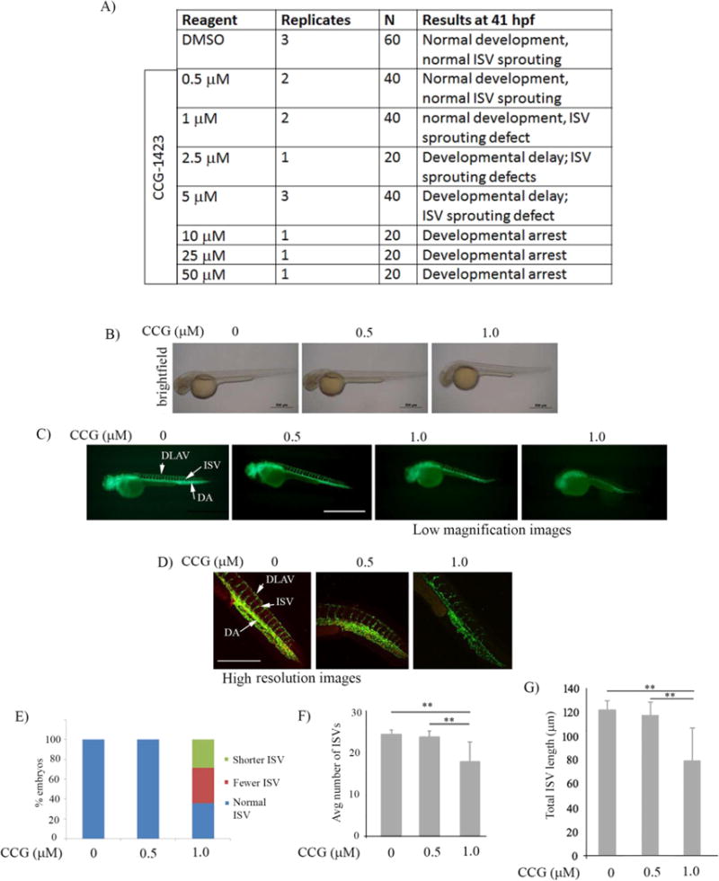
Figure 2. CCG-1423 inhibits developmental angiogenesis in zebrafish embryos.

A) Summary of phenotypes of Tg(flk1:gfp)lal16 zebrafish embryos at 22 hr after treatment with the indicated doses of either CCG-1423 or DMSO (control); N indicates the number of embryos in each treatment group. B-D) Brightfield (panel B; scale bar – 500 m) and fluorescence images (panels C [scale bar – 500 m] and D; panel D shows multiphoton images [scale bar – 50 m] of zebrafish embryos at the indicated doses of CCG-1423 relative to DMSO (ISV: intersegmental vessel; DA – dorsal aorta, DLAV – dorsal longitudinal anastomotic vessel). E-G) Bar graph summarizing the ISV phenotypes (panel E), average number of ISV (panel F) and the total length of ISV (panel G) per embryo at 41 hpf under DMSO vs low concentration CCG-1423 treatment (** p < .01).
