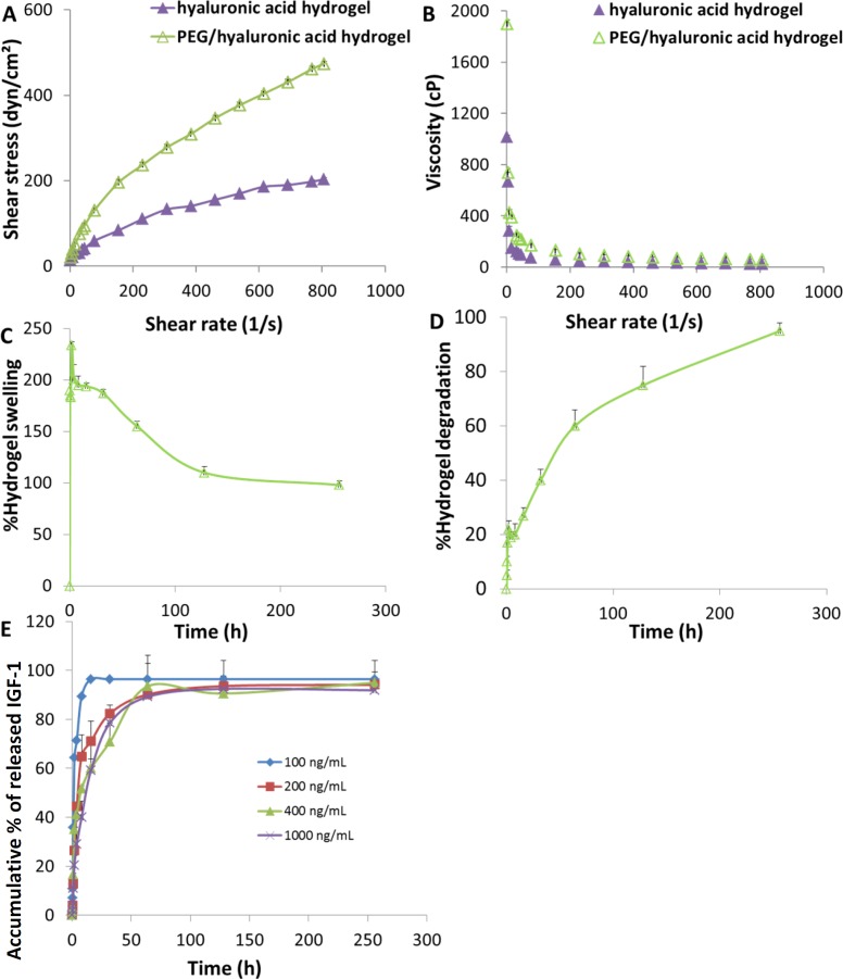
Figure 1.
Hydrogel scaffold characterization. (A) Rheological profiles, (B) viscosity curves; (C) swelling and (D) degradation behaviors of the PEG/hyaluronic acid scaffold incubated at 37 °C in PBS (pH 7.4). (E) Release profiles of IGF-1 from the scaffolds loaded with different cargo concentrations in PBS (pH 7.4) (n = 3, mean ± SD).

