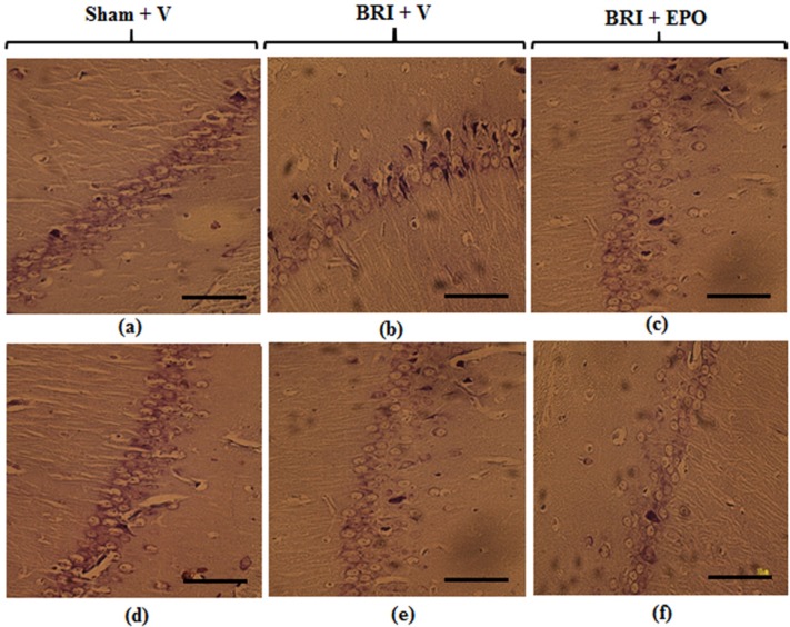
Figure 3.
Neurons of the CA1 region of hippocampus 24 h and 1w after reperfusion. Most neurons of the hippocampus in sham + V group have normal morphology (a) but for the BRI + V group, several degenerated cells can be seen with shrinkage nuclei and dark cytoplasm (b). EPO could attenuate degenerative changes induced by BRI (c). Normal morphology of pyramidal neurons was observed in all experimental groups (d_f) (Scale bars = 50 μm

