Figure 6.


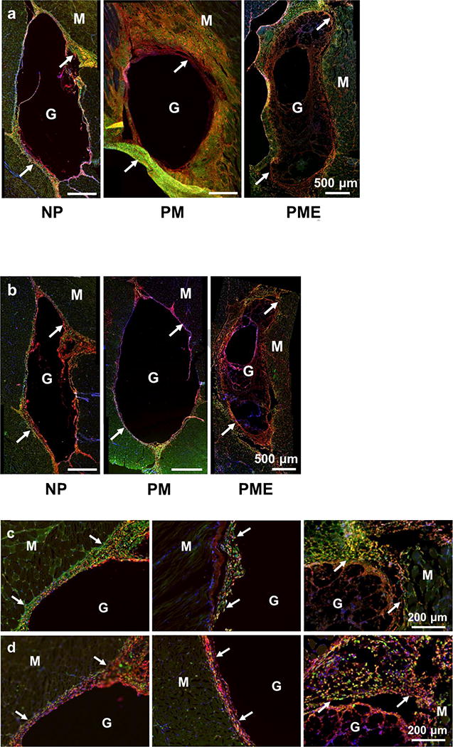
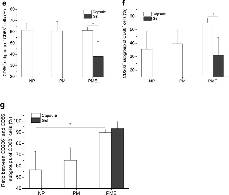
Macrophage polarization 3 days after hydrogel injection. M: muscle, G: hydrogel, Arrows: foreign body capsule. (a) CD86 (green) and CD68 (red) staining. (b) CD206 (green) and CD68 (red) staining. (c) CD86 (green)/CD68 (red) and (d) CD206 (green)/CD68 (red) staining at the hydrogel/muscle interface. (e, f) Percentage of CD86+ and CD206+ cells in CD68+ population, respectively. (g) Ratio between CD86+ and CD206+ cells in CD68+ population. * Significant difference, p < 0.05.
