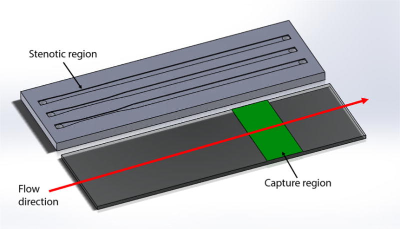
Figure 1.

Schematic diagram of a parallel flow assay. The upstream stenotic priming region was created by controlling the width of flow cells. Proteins were immobilized downstream on a reactive glass substrate by μCP to serve as platelet capture regions.
