
Figure 3.
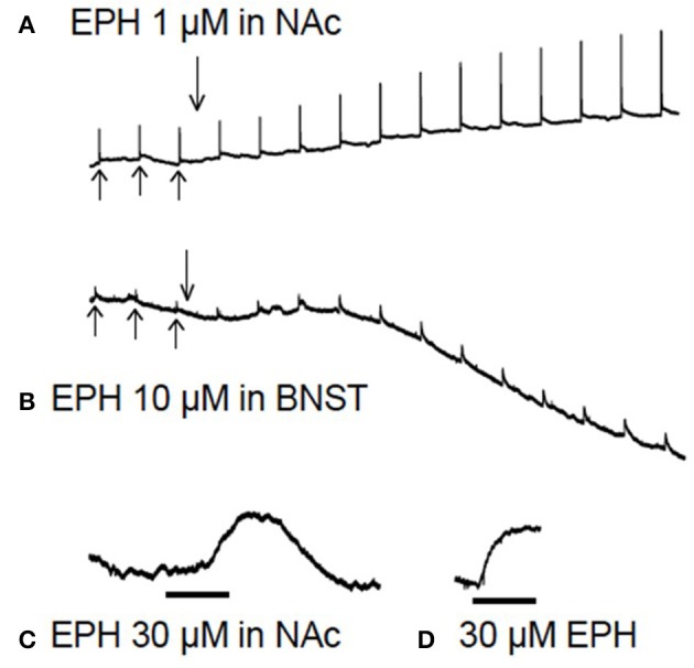
Raw data from brain slice experiments. In (A,B), after 3 baseline electrical stimulations (upward arrows), the drug is added (downward arrow) for 60 min while stimulations continue every 5 min. In the lower traces (C,D) there are no electrical stimulations. (A) Shows evoked dopamine efflux in the nucleus accumbens (NAc) after application of 1 μM ethylphenidate (EPH), note the increase in electrically evoked dopamine. (B) Application of EPH (10 μM) in the bed nucleus of the stria terminalis (BNST), note the increase in stimulated noradrenaline efflux and the apparent increase in background levels of transmitter 10 min after drug application. (C) Application of 30 μM EPH (black bar = 10 min) in the NAc appeared to increase basal dopamine levels (there is no electrical stimulation in this trace), but on testing 30 μM EPH at the carbon electrode in a calibration it was found that this drug is electroactive (D), although not nearly as electroactive as dopamine or noradrenaline.