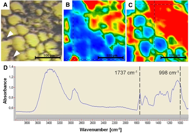
Fig. 5.
Light-microscopic (A) and FTIR images, of valerian root cross section showing high absorbance for mainly bornyl acetate at 1737 cm−1 (B) and cellulose at 998 cm−1 (C). The spectrum was taken from the cross mark in the appropriate FTIR images for cellulose (D). Colors blue, green, yellow, and red represent increasing content—the warmer the color, the higher the spectral intensity

