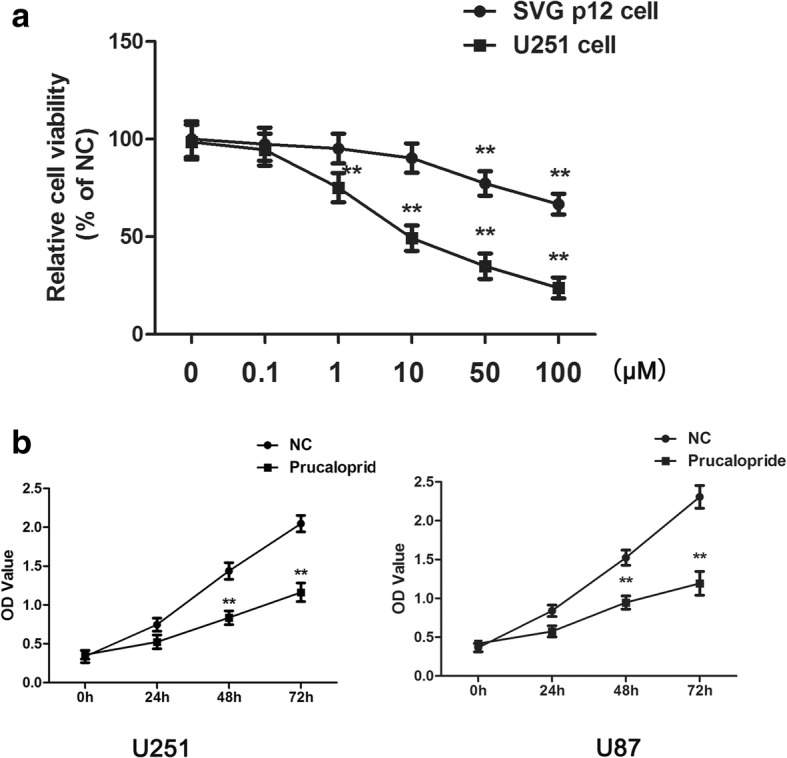
Fig. 1.

The suppressive effect of Prucalopride on glioma cells proliferation. a The viabilities of U251 and SVG p21 cells treated by Prucalopride with 0, 0.1, 1. 10, 50, 100 μM. **P < 0.01 versus control group (0 μM Prucalopride group). b The OD values of U251 and U87 cells treated by Prucalopride with 10 μM (the following experiments were performed according to the concentration) after 0, 24, 48 and 72 h. The data are presented as the means ± SD, **P < 0.01 versus NC group. Each assay was conducted in triplicate
