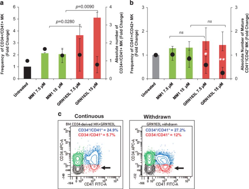
Figure 2.

Effects of imetelstat on the normal MK phenotype. (a) MK cultures that were untreated and treated with 7.5 and 15 μM of either MM1 or imetelstat (GRN163L) for 14 days were labeled with FITC-conjugated anti-CD41 and APC-conjugated anti-CD34 antibodies and analyzed by flow cytometry. The columns represent fold changes in the frequency (left y axis) and the absolute number (right y axis) of CD34+/CD41+ MK precursors quantified in the indicated culture conditions. (b) Cells from the same cultures as in panel (a) were labeled with FITC-conjugated anti-CD41 and APC-conjugated anti-CD42b antibodies and analyzed by flow cytometry. The columns represent fold changes in the frequency (left y axis) and the absolute number (right y axis) of mature CD41+/CD42b+ MK quantified in the indicated culture conditions. (c) Representative flow cytometric analyses of MK cultures treated continuously with imetelstat for 9 days (left panel) and of MK cultures in which imetelstat was removed after 6 days (right panel). Immature CD34+/CD41+ and more mature CD34−/CD41+ MK populations are indicated by blue and red colors, respectively. The results in panels (a and b) represent the mean ± s.d. of the frequency and corresponding absolute numbers detected in three independent experiments utilizing CD34+ cells from different healthy donors. NS, not statistically significant.
