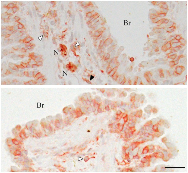
Figure 3.

Immunohistochemistry of the mouse lungs for CADM1. The lung sections were stained immunohistochemically with an anti-CADM1 antibody and counterstained with hematoxylin. Representative results are shown; upper, 30W-C and lower, 30W-Ex. White and black arrowheads indicate mast cells and a possible fibroblast, respectively. Br, bronchus; N, nerve. Bar = 25 μm.
