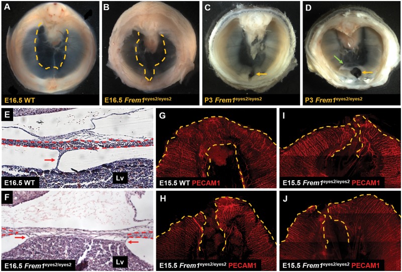
Figure 2.
Morphogenetic abnormalities associated with anterior sac hernia development in FREM1-deficient mice. (A, B) By E16.5 wild-type embryos on a mixed B6Brd/129S6 background showed complete muscularization of the anterior diaphragm. In contrast, Frem1eyes2/eyes2 embryos on an inbred B6Brd/129S6 background have a persistent region of amuscular diaphragm directly behind the sternum. Dashed yellow lines mark the boundary between amuscular and muscular regions of the diaphragm. (C, D) At P3 and P4, 45% (5/11) of Frem1eyes2/eyes2 embryos on an inbred B6Brd/129S6 background had a persistent region of amuscular diaphragm directly behind the sternum (yellow arrow in panel C) and 27% (3/11) had frank herniation with a sac. The hernia contents have been reduced in the embryo pictured in (D) leaving a hernia sac (green arrow) and a circular defect in the diaphragm (yellow arrow) that is completely surrounded by muscular diaphragm. (E, F) Coronal sections through a wild-type E16.5 embryo on a mixed B6Brd/129S6 background, reveal complete muscularization of the anterior diaphragm (dashed red lines). In these embryos, the liver was attached to the anterior diaphragm only by the falciform ligament (red arrow). In contrast, the muscular components of the diaphragm (dashed red lines) fail to meet in the midline in a subset of E16.5 Frem1eyes2/eyes2 embryos on an inbred B6Brd/129S6 background. In these embryos, the liver is also abnormally attached to the anterior diaphragm (red arrows). (G–J) Whole mount immunohistochemistry using a PECAM1 antibody, reveals a network of blood vessels surrounding the central tendon in wild-type E15.5 embryos on a mixed B6Brd/129S6 background (yellow dashed lines). In contrast, a poorly vascularized region of the anterior diaphragm (yellow dashed lines) was identified in 3/5 (60%) of Frem1eyes2/eyes2 embryos on an inbred B6Brd/129S6 background at E15.5.

