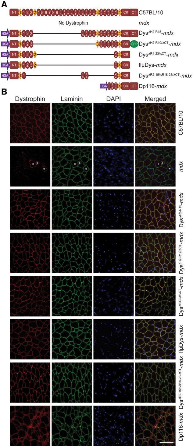
Figure 1.

Schematic representation of transgenic dystrophin constructs. (A) Full-length dystrophin in C57BL/10, absence of dystrophin in mdx and skeletal muscle-specific truncated dystrophins on the mdx background. HSA, human skeletal α-actin promoter; NT, N-terminus; diamonds represent hinge regions; ovals represent spectrin-like repeats; CR, cysteine-rich domain; CT, C-terminus. (B) Sarcolemmal localization of C57BL/10 and transgenic dystrophin. Dystrophin protein in mdx mice was present only in revertant fibers (denoted with *). Laminin outlines all individual fibers. Dystrophin (red), laminin (green), nuclei (DAPI). Scale bar is 100 µm.
