Figure 1.
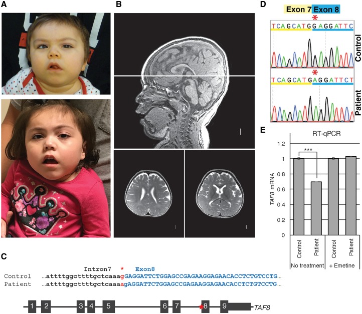
Developmentally delayed child identified with c.781–1G > A mutation in TAF8 gene. (A) Photographs of patient at 3 years of age (top) and 4 years of age (bottom). (B) Brain MRI acquired at 2 years and 7 months of age shows (sagittal on top and two transversal levels on bottom) mildly prominent extra-axial spaces, diffuse thinning of white matter with delayed myelinization, borderline enlarged lateral ventricles, short corpus callosum with narrow posterior body and absent splenium, normal brainstem, borderline small cerebellar vermis, and mildly small posterior fossa size. (C) (Top) Patient and control genomic sequences at the intron 7–exon 8 boundary of interest. Asterisk highlights the G > A splice site mutation (in red). Capital letters show exon 8 coding nucleotides. (Bottom) Schematic representation of the TAF8 gene (not to scale) with the position of the mutation indicated by the red asterisk. (D) Sequencing chromatogram highlighting the G nucleotide missing from the beginning of exon 8 in the patient cDNA. (E) RT-qPCR performed with primer annealing to the boundaries between exons 2 and 3, showing abundance of TAF8 mRNA in control and patient cells treated with and without emetine. Error bars represent SEM of three technical replicates. ***P < 0.001.

