Figure 3.
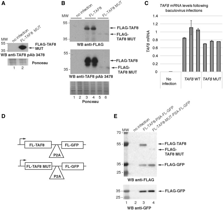
Exogenously expressed TAF8 mutant protein is unstable. (A) WCEs from insect Sf9 cells were prepared with either no infection, or with infection of a recombinant baculovirus encoding the TAF8: c.781-1G > A mutant protein fused to an FLAG tag and western blot assays were carried out using purified anti-TAF8 pAb 3478. (B) Western blot analyses of WCEs generated from Sf9 cells either with no infection, infection with recombinant baculovirus encoding FLAG tagged WT TAF8, or FLAG-tagged mutant TAF8 (FL-TAF8 MUT) protein, probed either with anti-FLAG antibody (upper blot), or affinity purified anti-TAF8 pAb 3478 (middle blot). Each lane represents an independent infection. (C) RT-qPCR performed with primers annealing to the boundary between exons 2 and 3 of TAF8 mRNA normalized to Sf9 housekeeping genes 28S, GAPDH and L35, performed on cDNA generated from Sf9 cells with either no infection, or infected with recombinant baculovirus encoding WT TAF8 protein or TAF8: c.781-1G > A mutant protein. All values were normalized to the average of the WT TAF8 infection. Error bars represent SEM of three technical replicates. See Supplementary Material, Table S1 for primer sequences. (D) Schematic representations of the cassettes expressing a single mRNA encoding either WT FLAG-TAF8 or mutant FLAG-TAF8 together with FLAG-GFP separated by the self-cleavage P2A sequence. (E) Western blot analyses of WCEs generated from Sf9 cells either with no infection, infection with recombinant baculovirus encoding FLAG tagged WT TAF8-P2A-FL-GFP, or FLAG-tagged mutant TAF8 (FL-TAF8 MUT- P2A-FL-GFP) protein, probed either with anti-FLAG antibody (upper blot), anti-GFP (lower blot). In A and B, for Ponceau staining 5× the amount of protein was loaded as compared with the western blots. In A, B and E, the molecular weight (MW) markers are indicated in kDa.

