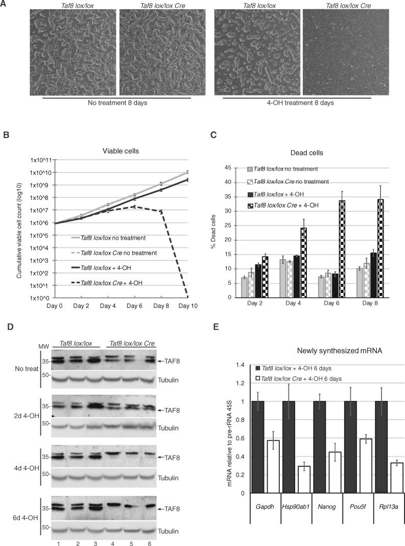
Figure 4.
mESCs induced to delete Taf8 undergo cell death potentially as a result of transcriptional defects. (A) Representative images of Taf8lox/lox or Taf8lox/lox-Rosa26-CreERT2 mESCs after 8 days of culture with or without 0.5 µM 4-hydroxy tamoxifen treatment (4-OH). See also Supplementary Material, Figure S2A. (B) Curve showing the cumulative number of viable mESCs over the course of the treatment, cell counts were plotted taking into account the dilution factor at each passage. (C) Cell death was measured by trypan blue exclusion over the course of the treatment (as indicated). (D) Western blot analyses to detect TAF8 protein with the affinity purified 3478 pAb during several days (d) of 4-hydroxy tamoxifen (4-OH) treatment. Equal loading was measured by using an anti-γ-tubulin antibody. MW markers are indicated in kDa. (E) RT-qPCR of randomly selected, highly expressed genes, measuring newly synthesized mRNA relative to Pol I transcribed pre-rRNA 45S. The list of the primers is indicated in Supplementary Material, Table S1. For all experiments three biological replicates were carried out per genotype. Error bars represent SEM.

