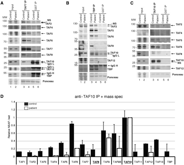
Figure 5.
TFIID assembly is defective in fibroblasts harboring the TAF8: c.781–1G > A mutation. (A–C) Coimmunoprecipitation performed on WCEs prepared from control, or TAF8: c.781-1G > A patient fibroblasts, using either anti-TAF7 and anti-GST (negative control) antibodies (A) or anti-TBP and anti-GST (B) or anti-TAF10 and anti-GST (C). Input extracts or immunopurified complexes were separated by SDS-PAGE and western blot analyses were carried out by probing the blots with antibodies against the indicated TFIID subunits. The anti-GST antibody heavy (IgG H) and light chains (IgG L) are indicated. Ponceau-stained membranes in A–C show equal loading. In A–C the MW markers are indicated in kDa. (D) Mass spectroscopy performed on proteins co-IP-ed with anti-TAF10 antibody, normalized to TAF10 bait protein. Relative NSAFbait was calculated as described previously (52). Error bars represent SEM of two technical replicates. See Supplementary Material, Table S2 for raw mass spectrometry peptide counts.

