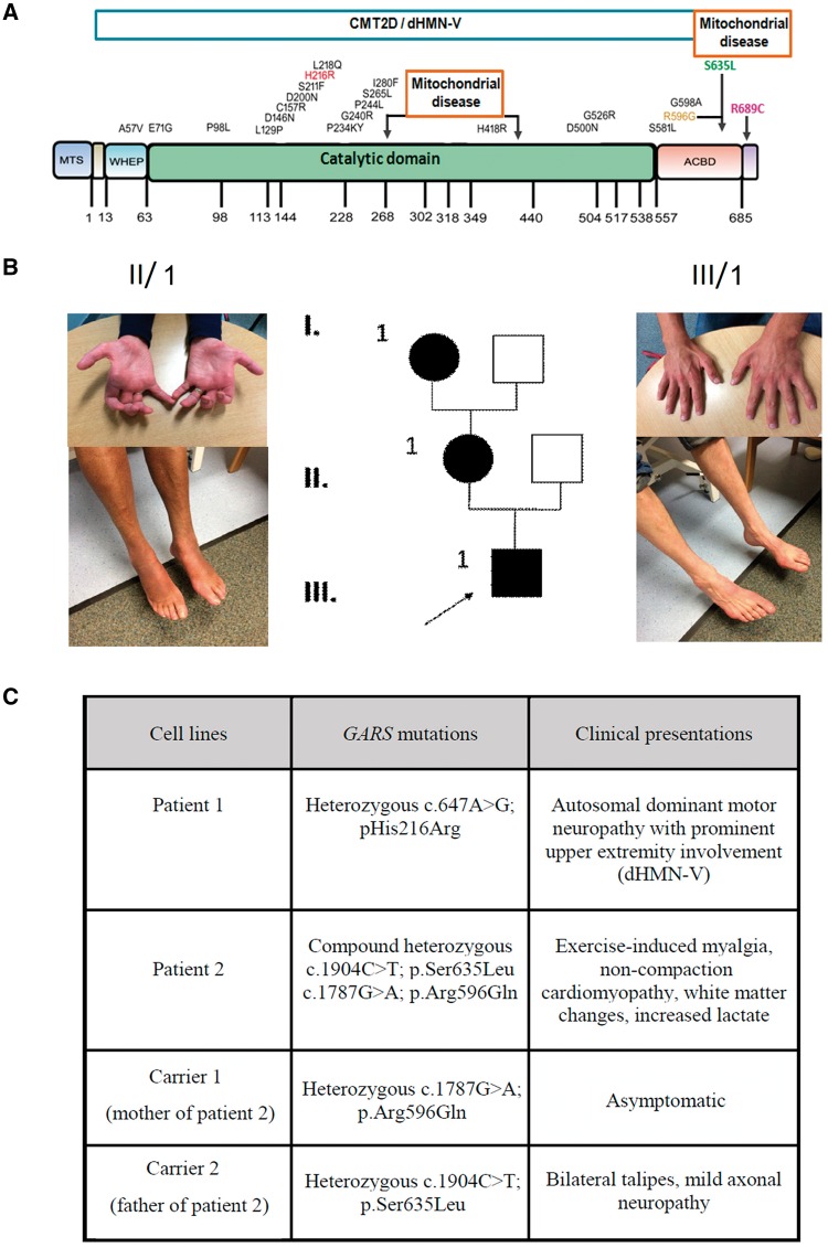
Figure 1.
Schematic representation of the GARS protein and distribution of dHMN-V and mitochondrial disease-associated dominant and recessive mutations. (A) Dominant mutations causing CMT2D/dHMN-V are mostly located in the catalytic domain marked with black. Recessive mutations leading to mitochondrial disease localized in the catalytic domain and at the anticodon binding domain (ACBD) shown by the black arrows. Mutations modelled in this study are highlighted with red, orange, green and purple. (B) Pedigree of patient 1 with a novel heterozygous c.647A>G, p.(His216Arg) mutation, both patient 1 and his affected mother show prominent atrophy of small hand muscles and moderate atrophy and weakness in the feet. (C) Summary of the clinical presentations of the patients (patient 1 and 2) and heterozygous parents of patient 2 (carrier 1 and 2) whose fibroblasts were used in this study.

