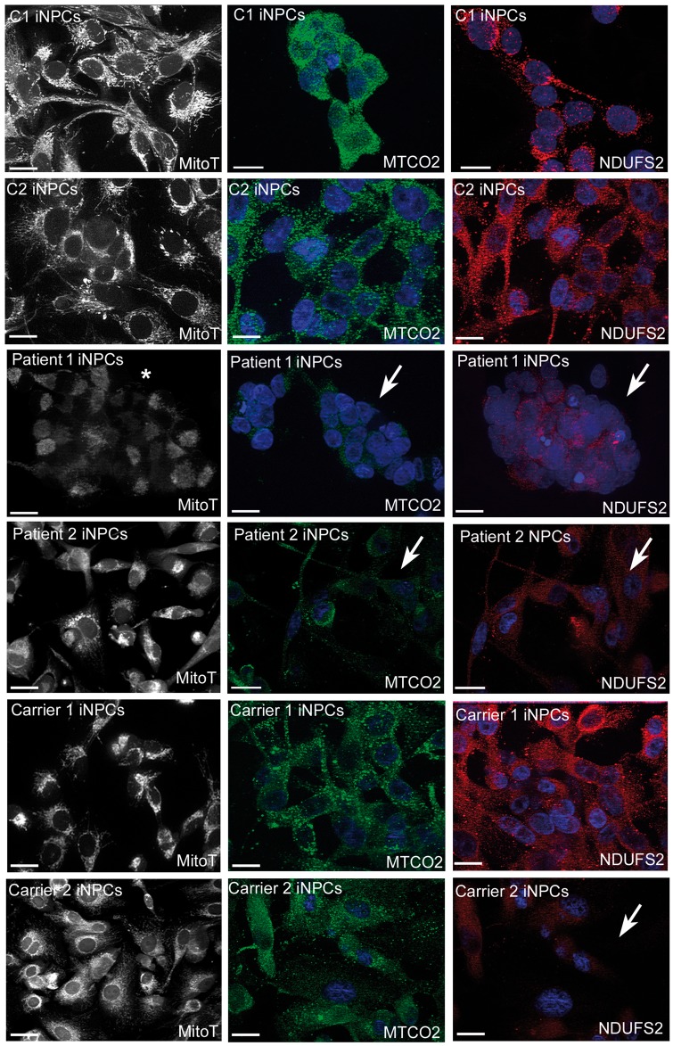
Figure 6.
Immunostaining of mitochondrial proteins in human fibroblasts and iNPCs. Representative fluorescent images of mitotracker (black and white), MTCO2 (green channel) and NDUFS2 (red channel). Abnormal MitoTracker staining indicates abnormal mitochondrial membrane potential in patient derived iNPCs (marked with white star). iNPCs with GARS mutations in patient 1, patient 2 and carrier 2 show weak signals of mitochondrial proteins (indicated with white arrows) confirming mitochondrial translation defect (scale bar represents 20 um).

