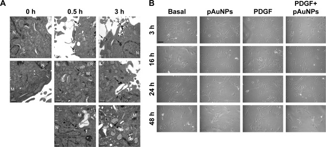
Figure 2.
TEM and morphological analysis of VSMCs treated with pAuNPs.
Notes: VSMCs were incubated with pAuNPs for the indicated time intervals. (A) The subcellular distribution of the pAuNPs was analyzed by TEM. Scale bar =0.5 μm. (B) Cell morphology was analyzed by phase-contrast microscopy. Scale bar =100 μm.
Abbreviations: TEM, transmission electron microscopy; pAuNPs, physically synthesized gold nanoparticles; VSMC, vascular smooth muscle cell; ER, endoplasmic reticulum; M, mitochondria; N, nucleus.

