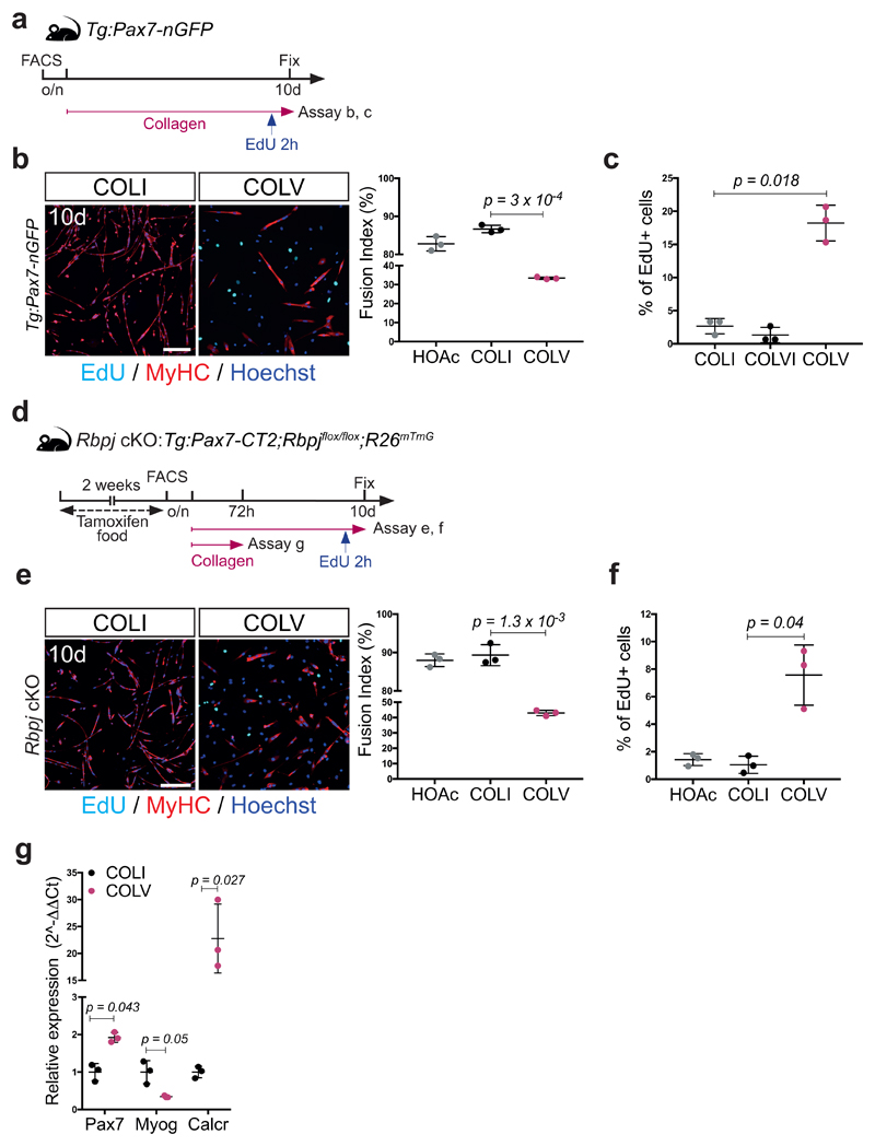
Extended data Figure 3. Collagen V delays proliferation and differentiation of satellite cells.
(a) Experimental scheme: isolated Tg:Pax7-nGFP satellite cells cultured overnight (o/n) before collagen treatment.
(b) Myosin heavy chain (MyHC)/EdU staining of satellite cells treated with COLI or COLV. Fusion index: 82%, 86%, and 33% for HOAc solvent, COLI, and COLV, respectively (n=3 mice, ≥250 cells, 2 wells/condition).
(c) Percentage of EdU+ primary myogenic cells after 10 days of culture with indicated collagens. EdU: 2.6%, 1.3% and 18.2% for COLI, COLVI and COLV respectively (n=3 mice, ≥250 cells, 2 wells/condition).
(d) Experimental scheme for Control and conditional knock-out mice. Satellite cells were plated overnight (o/n) before collagen treatment.
(e) GFP/Myogenin immunostaining of Control (Ctr) and Rbpj cKO satellite cells (n=3 mice/condition) incubated 60h in presence of COLI or COLV (n=3 mice, ≥200 cells, 2 wells/condition).
(f) Percentage of EdU+ cells (2h pulse) of Rbpj null primary myogenic cells, after 10 days of culture with HOAc or indicated collagens. EdU: 1.0 % and 7.6% for COLI and COLV respectively (n=3 mice, ≥150 cells, 2 wells/condition).
(g) RT-qPCR on GFP+ Rbpj null satellite cells isolated by FACS and cultured for 72h in the presence of COLI or COLV. Results are normalized to Tbp.
Error bars, mean ± SD; two-sided paired t-test; #p-value: two-sided unpaired t-test. Scale bar: 50μm.

