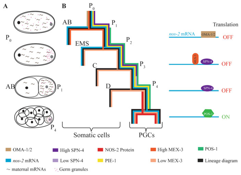
Fig. 6.2.
Translational control during germ cell specification in C. elegans. (a) Schematic representation of asymmetric cleavage and asymmetric distribution of maternal components. Pink dots and tilde-like structures represent the germ granules and the maternal mRNAs, respectively. (b) Distribution patterns of RBPs and nos-2 mRNA during early embryonic cleavages; colors representing the different components are indicated at the bottom. Translation of nos-2 mRNA is suppressed sequentially by OMA-1 and OMA-2 in oocytes (not shown here), by MEX-3 in the AB blastomere, and by SPN-4 in the P lineage until P3. The rapid decrease in the SPN-4 to POS-1 ratio in P4 enables POS-1 to compete out SPN-4 for binding to the nos-2 3′ UTR, which depresses nos-2 translation in P4

