
Fig. 12.
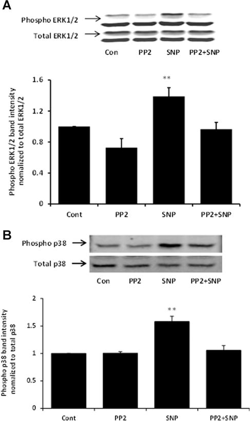
Western blots showing the inhibitory effect of PP2 on ERK1/2 (Part A) and p38 (Part B) phosphorylation in cells exposed to 100 μM SNP. The samples were obtained from cells that had been pre-incubated with PP2 (10 μM) for 40 min before being exposed to SNP for 10 min in the continued presence of PP2. In each part, a typical Western blot (upper) together with a bar graph (lower) showing the band density results (mean ±SEM), pooled from three independent experiments, are presented. **Indicates a significant difference from control, P < 0.01.