
Fig. 5.
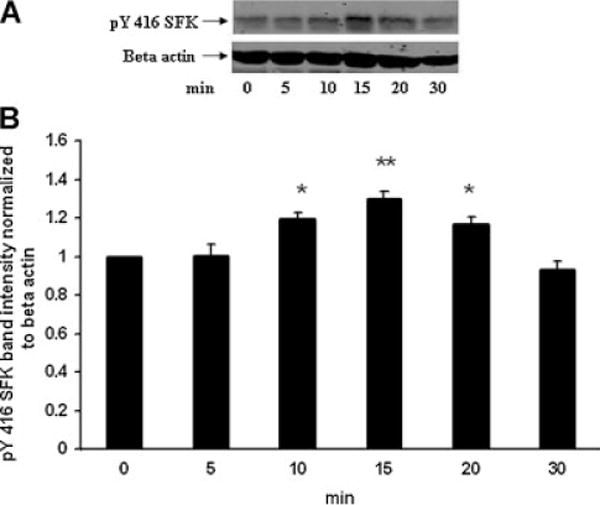
Time course showing changes of SFK phosphorylation detected in the epithelium removed from intact porcine lenses following incubation for 5–30 min in the presence of ET-1 (100 nM). The control group (time 0) received no ET-1. Part A: A Western blot showing pY416 SFK-immunoreactive band density. β-actin served as a loading control. Part B: Pooled data on pY416 SFK band density relative to β-actin band density. The values are the mean ± SE of results from three lenses. *P < 0.05 and **P < 0.01 indicates a significant difference from control.