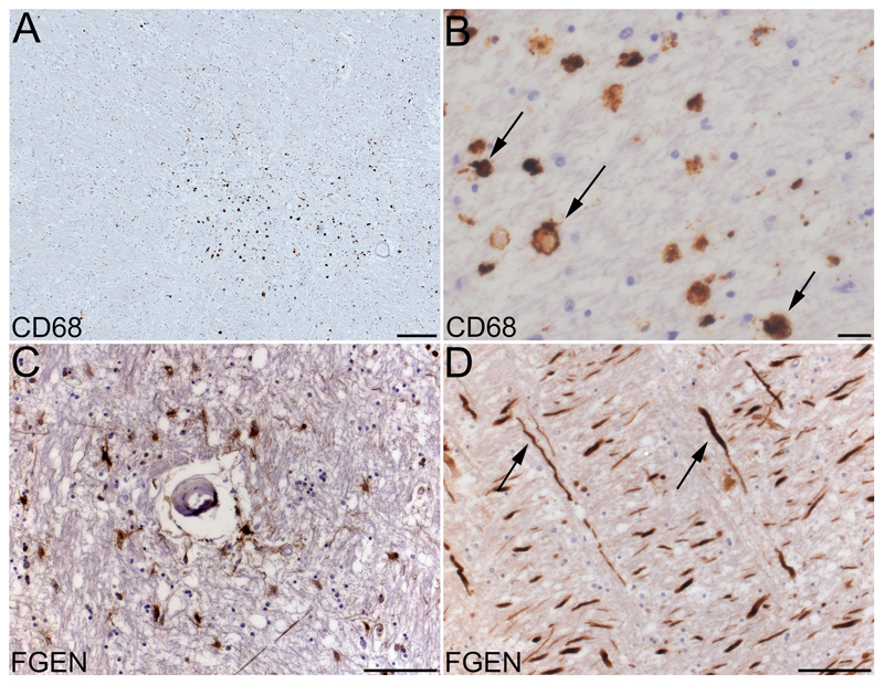
Figure 1.
Histopathology of white matter lesions and BBB dysfunction in subcortical white matter. A, CD68-PGM1 immunolabelled section shows a histopathological white matter lesion, defined by a cluster of amoeboid CD68 positive cells. B, CD68 positive amoeboid cells, assumed to be phagocytic microglia (arrows show examples) at higher magnification. C, fibrinogen positive cells and axons, around a small blood vessel. D, nerve axons strongly positive for fibrinogen (arrows show examples). Scale bars 200 µm (A), 20 µm (B) or 100 µm (C, D).

