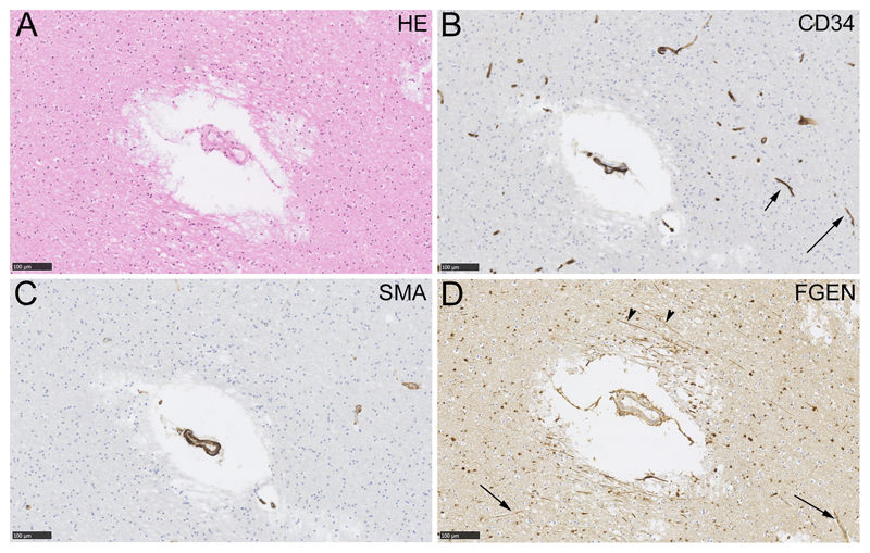
Figure 2.
Fibrinogen immunolabelling around a small artery in deep subcortical white matter. Neighbouring sections were labelled with haematoxylin and eosin (HE, panel A), an endothelial marker (CD34, panel B), a myocyte marker (smooth muscle action, SMA, panel C) or fibrinogen (FGEN, panel D). Capillaries are evident in the CD34-labelled section (arrows, B) and not in the SMA-labelled section. In panel D, fibrinogen labelling is evident around the small artery in cells and in axons (arrowheads). In this example, enhanced perivascular fibrinogen labelling is not seen around capillaries (arrows). Scale bars 100 µm.

