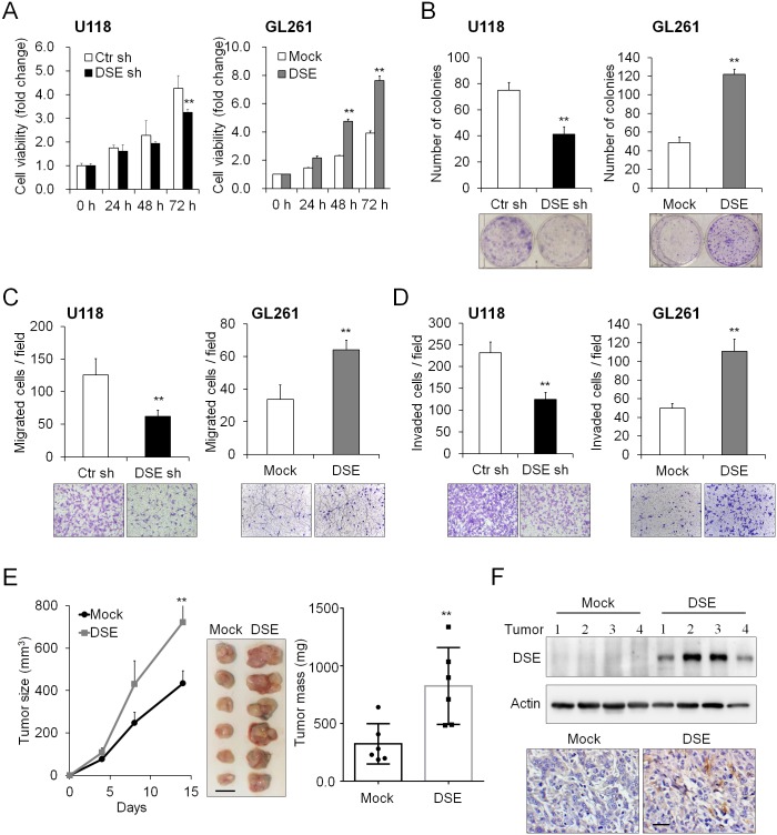
Fig 3. Dermatan sulfate epimerase 1 (DSE) regulates malignant phenotypes in glioma cells.
(A) DSE modulated cell viability in vitro. The cell viability of U118 and GL261 cells was determined using a CCK-8 assay at the indicted time-points. Data represent means ± SD from three independent experiments. *P < 0.05; **P < 0.01. (B) Effects of DSE on anchorage-dependent colony formation. Representative images of colonies are shown at the bottom. Results are presented as the mean ± SD from three independent experiments. **P < 0.01. (C) Effects of DSE on Transwell cell migration, and (D) Matrigel invasion. Representative images are shown at the bottom. All results are represented as means ± SD from three independent experiments. **P < 0.01. (E) DSE enhanced tumor growth in vivo. GL261 transfectants were subcutaneously injected into C57BL/6 mice. The size of the tumors was measured at the indicated time-points, and is represented as the mean ± SD. Tumors were excised and weighted on the 14th day. **P < 0.01, n = 6. Scale bars, 0.5 cm. (F) Expression of DSE in excised tumors. The protein lysate was analyzed by western blotting (top). Actin was used as a loading control. Immunohistochemistry of DSE in tumors (bottom). Representative images are shown. Scale bars, 50 μm.

