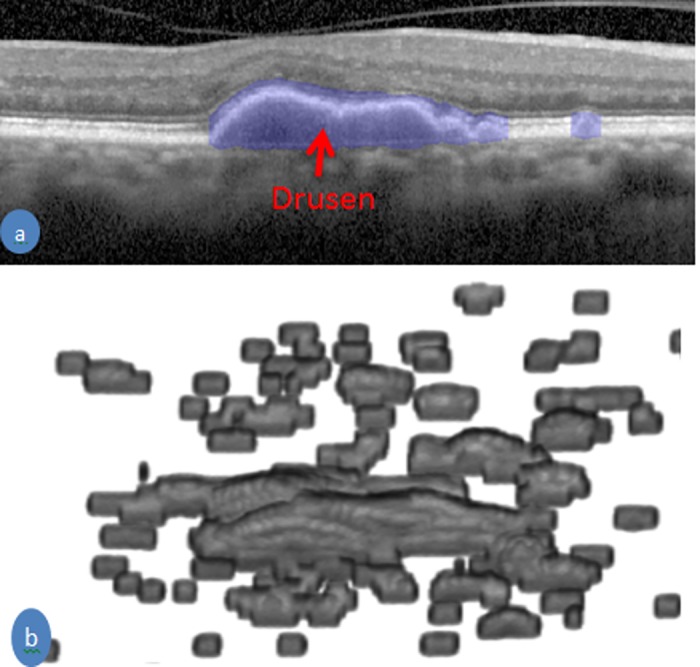
Fig 6. An example of drusen segmentation.

(a) an SD-OCT B-scan with delineation of drusen by the blue color (b) Drusen in 3D view of an SD-OCT volume of an AMD patient.

(a) an SD-OCT B-scan with delineation of drusen by the blue color (b) Drusen in 3D view of an SD-OCT volume of an AMD patient.