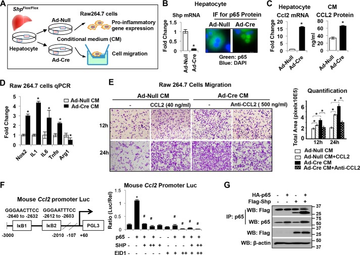
Figure 7.
Deletion of Shp in hepatocytes increases CCL2 production leading to macrophage proinflammatory polarization. A, schematic diagram shows experimental design. Primary hepatocytes from Shpflox/flox mice were infected with adenovirus expressing Cre recombinase (Ad-Cre) or vector control (Ad-Null). CM was collected at 24 h post-adenoviral vector infection and used for RAW cell treatment. B, left, relative Shp mRNA levels were determined by qPCR. Data are represented as mean ± S.D. for triplicate experiments/group; *, p < 0.05. Right, representative images of immunofluorescence (IF) staining of p65 in hepatocytes. C, left, qPCR analysis of relative Ccl2 mRNA levels in hepatocytes. Right, CCL2 protein level in CM measured by ELISA. Data are represented as mean ± S.D. for experiments/group; *p < 0.05. D, RAW cells were incubated with CM from hepatocyte culture for 6 h, and the relative expression of genes involved in inflammation was determined by qPCR. Data are represented as mean ± S.D. for triplicate experiments/group; *, p < 0.05. E, left, representative images of RAW cell migration. RAW cells were incubated with CM in the presence or absence of recombinant mouse CCL2 (40 ng/ml) or anti-mouse CCL2 antibody (500 ng/ml). Cell migration was assessed after incubation for 12 and 24 h, respectively. Right, quantitation of cell migration was determined by measuring the pixel density of crystal violet–stained cells using ImageJ software. Data are represented as mean ± S.D. for five fields/sample. *, p < 0.05. F, left, diagram shows the location of two IκB sites on the mouse Ccl2 promoter reporter (Ccl2-Luc). Right, AML12 cells were transfected with Ccl2-Luc with various expression plasmids. Luciferase activities were determined at 24 h post-plasmid transfection. Data are calculated as the ratio of firefly luminescence divided by Renilla luminescence and presented as mean ± S.D. for triplicate experiments/group. *, p < 0.05. G, AML12 cells were overexpressed with FLAG-SHP or HA-p65 and harvested at 24 h post-plasmid transfection. Immunoprecipitation (IP) followed by Western blotting (WB) was employed to detect protein-protein interactions between FLAG-SHP and HA-p65.

