Figure 2.
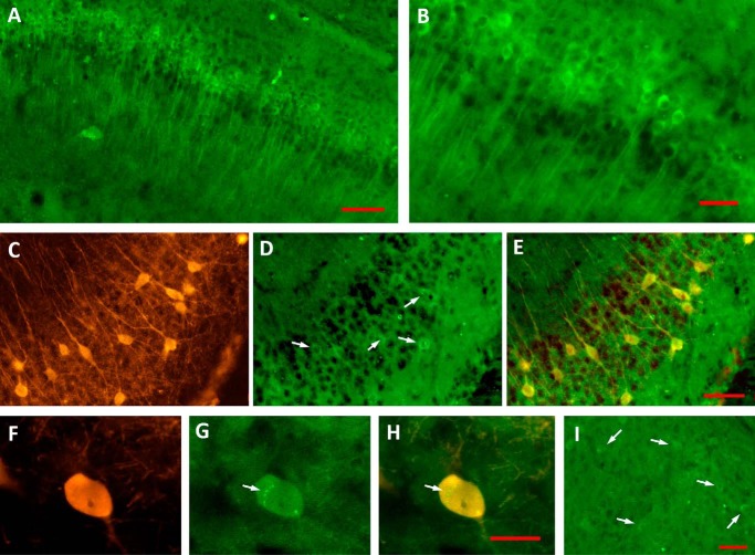
Tau phosphorylation was found increased in Tau N-terminal domains. A and B, the increase of Tau phosphorylation in 3xTg-AD group at sites labeled by mAb AT180 was found coexisting with the pyramidal neurons from the CA1/subiculum (A). A magnification is shown in B. C–E, PARV+ hippocampal interneurons (C) accumulate phosphorylated forms of Tau (D and E, white arrows). F–H, AT180–positive PARV+ interneurons (F) revealed punctate accumulation of phospho-Tau (G and H, white arrows). Entorhinal neurons displayed lower levels of phospho-Tau (I, white arrows). Scale bar for A, 100 μm; scale bars for B–E, 50 μm; scale bar for F–H, 20 μm; and scale bar for I, 50 μm.

