Figure 4.
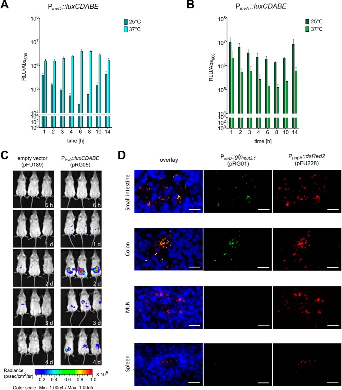
Expression analysis of InvD in vitro and in vivo. A and B, expression of InvD and InvA under in vitro conditions. Overnight culture of Y. pseudotuberculosis YPIII pRG05 (PinvD::luxCDABE) was diluted 1:50 and grown at 25 or 37 °C in LB medium. Light emission of the luxCDABE reporter and optical density were measured. The dashed line represents the detection limit. C, in vivo expression analysis of an invD-luxCDABE fusion shows expression of invD during infection. BALB/c mice were orally infected with 9 × 10⁸ CFU of Y. pseudotuberculosis YPIII pFU189 (empty vector) and YPIII pRG05 (PinvD::luxCDABE). Mice were anesthetized and ventrally imaged with the in vivo imaging system camera at given time points. D, expression of invD in murine intestines visualized with GFP reporter fusion. BALB/c mice were infected orally with 3 × 10⁸ CFU of Y. pseudotuberculosis YPIII pFU228/pRG01 (PgapA::dsred, PinvD::gfpmut3.1). Mice were sacrificed 3 days post-infection, and the small intestine, colon, mLNs, and spleen were isolated. Cryosections (thickness, 7 μm) were prepared and imaged with a fluorescence microscope. Bacteria were detected by the DsRed2 reporter protein. Cell nuclei were stained with DAPI. White bars indicate 10 μm.

