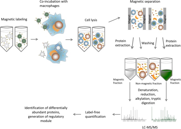
Fig. 1.
Flowchart for the purification of conidia-containing phagolysosomes, proteome detection and data analysis. The procedure starts with magnetic labeling of conidia that are then incubated with macrophages for 2 h. The cell lysate is loaded onto a magnetic column to separate the magnetic fraction from the cell debris. The protein is extracted on-column, then precipitated and concentrated for LC-MS/MS measurement. After label-free quantification the data was analyzed to identify regulated proteins and modules.

