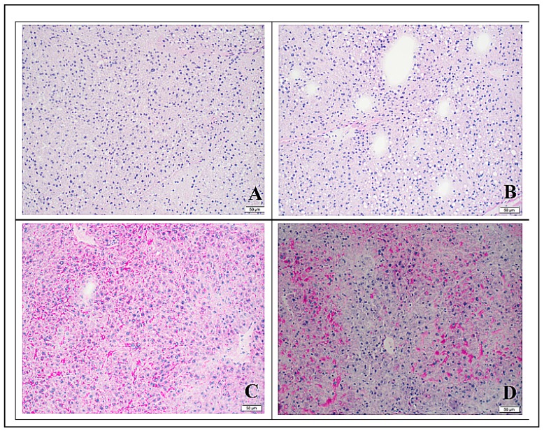
Figure 3.
Periodic acid Schiff (PAS) stained liver tissue (20×, scale bar represents 20 µm) collected from mice fed maize vegetable diet (MVD) diets or chow. Bright red staining areas indicate the presence of polysaccharides; dark blue staining indicates nuclear material. (A) Liver section from mouse fed the unsupplemented MVD for 6 days; (B) liver section from mouse fed the unsupplemented MVD for 13 days; (C) liver section from mouse fed the MVD supplemented with choline for 9 days; and (D) liver section from a control mouse fed chow for 13 days.

