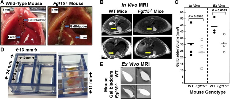
Figure 1. Altered gallbladder shape and diminished gallbladder filling in FGF15-deficient mice.
(A) Photographs of WT and FGF15-deficient mouse gallbladders in situ at laparotomy. To improve visual clarity, dashed lines demarcate gallbladders. FGF15-deficient mouse gallbladders were tubular in appearance compared to the spherical gallbladders observed in WT mice. Size bars, 1 mm. (B) Representative images from in vivo MRI of WT and FGF15-deficient mice show differences in gallbladder shape. Arrows demarcate gallbladders. (C) In vivo (left) and ex vivo (right) measurements of volume of gallbladders from WT and FGF15-deficient mice. Each symbol represents one mouse gallbladder. Horizontal lines demarcate mean values. Mean ex vivo volumes were significantly greater in gallbladders from WT compared to FGF15-deficient mice; P=0.0399). (D) Septate Lucite box used for ex vivo imaging of murine gallbladders. Measurements indicate internal and external dimensions of tray containing 2% agarose. Right panel show vertical dimension and gallbladder surrounded by 2% agarose in chamber. (E) Representative images from ex vivo MRI of gallbladders from WT and FGF15-deficient mouse highlight differences in gallbladder shape.

