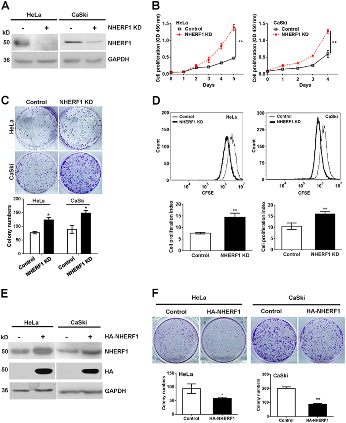
Fig. 2. NHERF1 inhibits cervical cancer cell proliferation.
a Knockdown of NHERF1 expression in cervical cancer cells was verified by immunoblotting analysis. HeLa cells were stably transfected with shNHERF1 constructs (HeLa-NHERF1-KD), and CaSki cells were transiently transfected with NHERF1 siRNAs (CaSki-NHERF1-KD). b Knockdown of NHERF1 enhanced proliferation of cervical cancer cells. Proliferation of HeLa-NHERF1-KD, CaSki-NHERF1-KD, and their control cells was detected by CCK-8 at the indicated time points (repeated-measures analysis of variance, **p < 0.01, error bars represent mean ± s.d., n = 3). c Knockdown of NHERF1 enhanced the colony formation of cervical cancer cells. Colony formation was monitored in HeLa or CaSki cells for 7 days. Top panel: Representative photographs of the clonogenicity. Bottom panel: Quantification of the colony formation efficiency (t test, *p < 0.05, error bars represent mean ± s.d., n = 3). d Inhibition of NHERF1 expression enhanced cell proliferation of cervical cancer cells by CFSE assay (t test, **p < 0.01, error bars represent mean ± s.d., n = 3). Cells were stained with CFSE and analyzed following the protocol as described in the “Methods”. e Overexpression of NHERF1 in cervical cancer cells was verified by immunoblotting analysis. HeLa and CaSki cells were transiently transfected with NHERF1 constructs, respectively, and expression of NHERF1 was verified by western blotting. f Exogenous NHERF1 expression inhibited the colony formation of cervical cancer cells. Colony formation was monitored in HeLa or CaSki cells for 7 days. Top panel: Representative photographs of the clonogenicity. Bottom panel: Quantification of the efficiency of colony formation (t test, *p < 0.05, error bars represent mean ± s.d., n = 3). Cells proliferation was detected by CCK-8 assay at the indicated time points (repeated-measures analysis of variance, **p < 0.01, error bars represent mean ± s.d., n = 3)

