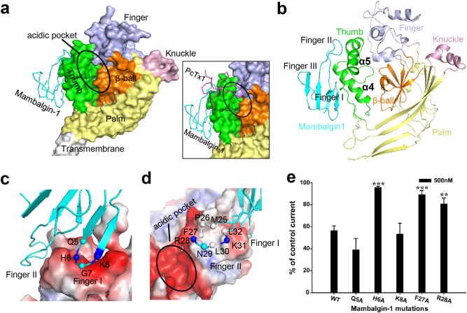
Fig. 3. Location and interaction of mambalgin-1 on cASIC1a.
a Location of mambalgin-1 in cASIC1aΔNC-mambalgin-1 complex. A single subunit of cASIC1a channel is shown in surface representation with each domain in a different color. Mambalgin-1 is shown in ribbon representation. Insert, the location of mambalgin-1 (cyan) overlapping with PcTx1 (magenta). The PcTx1 position was taken from the structure of PcTx1-bound cASIC1a (PDB 4FZ1). b Cartoon representation of a single subunit derived from the cASIC1aΔNC-mambalgin-1 complex. c, d Close-up views of the interactions of the Finger I region (c) and Finger II region (d) of mambalgin-1 with the thumb domain of cASIC1aΔNC. The Cα atoms of several key residues in the finger regions are shown as spheres. Basic residues of mambalgin-1 fingers are colored in blue, and the hydrophobic residues in white. e Bar graph representing the effect of different mambalgin-1 point mutants (500 nM) on wild-type cASIC1a. Data are means±S.E. (error bars) (*P < 0.05; **P < 0.01; ***P < 0.001; different from WT; t-test; n = 4–13)

