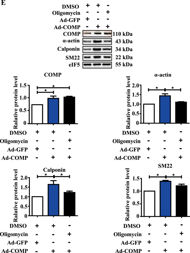
Fig. 5. Mitochondrial bioenergetics defects by COMP deficiency contributes to VSMC dedifferentiation.
a Traces and quantification of the mitochondrial oxygen consumption rates of serum-starved VSMCs stimulated with PGDF-BB (25 µg/L) for 48 h. The data were analyzed using unpaired two-tailed Student’s t test and presented as the means ± SD of three independent experiments. *P < 0.05. b Traces and quantification of the mitochondrial oxygen consumption rates of VSMCs stimulated with TGF-β (2.5 µg/L) for 48 h. The data were analyzed using unpaired two-tailed Student’s t test and presented as the means ± SD of four independent experiments. *P < 0.05. c Western blot analysis and quantification of the protein levels of α-actin, calponin and SM22 in VSMC cell lysates upon oligomycin (1 µM, 2 µM) stimulation. The data was analyzed using one-way ANOVA followed by the Student–Newman–Keuls test for post-hoc comparison and presented as the means ± SD of six independent experiments. *P < 0.05. d Western blot analysis and quantification of the protein levels of α-actin, calponin and SM22 in VSMC cell lysates upon transfection of vector or PGC1-α plasmid. The data were analyzed using paired two-tailed Student’s t test and presented as the means ± SD of three independent experiments. *P < 0.05. e Western blot analysis and quantification of the protein levels of COMP, α-actin, calponin and SM22 in VSMC cell lysates infected with COMP adenovirus or COMP adenovirus plus addition of oligomycin (2 µM). The data was analyzed using one-way ANOVA followed by the Student–Newman–Keuls test for post-hoc comparison and presented as the means ± SD of three independent experiments. *P < 0.05


