Figure 1.
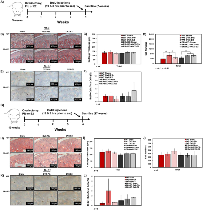
Role of estradiol via ERα on histomorphometry and cell proliferation of mandibular condylar fibrocartilage from 7-week and 17-week old WT and ERαKO female mice after sham, ovariectomy plus placebo treatment, and ovariectomy plus estradiol treatment. Data from 7-week old mice: (A) Timeline for experiment (B) Representative hematoxylin and eosin images of all groups. (C) Cartilage thickness and (D). Cell numbers determined by histomorphometry. (E) Representative BrdU images illustrating cell proliferation and (F). Quantification of percentage of BrdU+ cells. Data from 17-week old mice. (G) Timeline for experiment. (H) Representative hematoxylin and eosin images of all groups (I) Cartilage thickness and (J). Cell numbers determined by histomorphometry. (K) Representative BrdU images illustrating cell proliferation and (L). Quantification of percentage of BrdU+ cells. All values represent means ± standard deviation. n = 6 for all data, ^p < 0.01.

