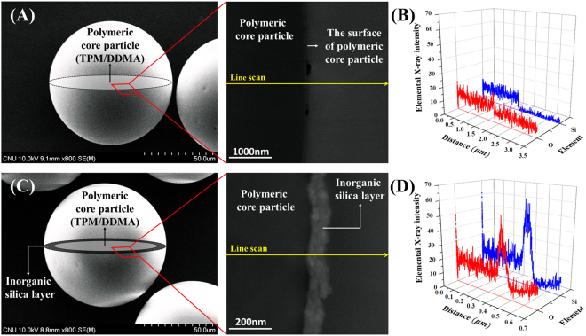
Figure 5.
Analysis of the core-shell structure in the particles. (A) SEM and TEM image of the polymeric core microparticles before the sol-gel reaction and (B) the corresponding line scanning profile of EDS along the cross section of the particles; (C) SEM and TEM image of the organic-inorganic hybrid particles after the sol-gel reaction and (D) the corresponding line scanning profile of EDS along the cross section of the particles. The SEM images are combined with the schematic illustrations of the expected structure. The enlarged TEM image of the cross section of the particle clearly indicates the existence of a shell layer. In the analysis of the line scanning profile of EDS, the blue and red line represent silicon and oxygen atoms, respectively.

