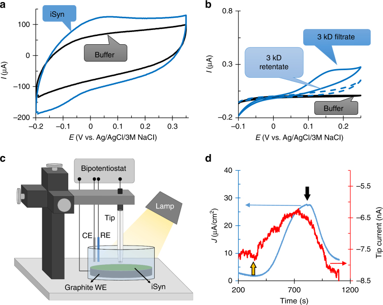
Fig. 5.
iSyn electron transfer to the BPEC is via a diffusive endogenous mediator. a CV in the light for buffer (black) and the iSyn (blue) separated by a 3 kD dialysis membrane on the graphite electrode. b CV of supernatant fraction from centrifuged iSyn cells separated by filtration through 3 kD cut-off centricon. Filtrate (solid blue line), retentate (dashed blue), and buffer (black). c Illustration of the scanning electrochemical microscopy setup. iSyn settled on the working electrode (WE) electrode. The counter electrode (CE) is platinum, and the reference electrode (RE) a Ag/AgCl/3 M KCl. The tip is a carbon-based microelectrode with a mixture of bilirubin oxidase (BOD) embedded in an Os-complex modified polymer matrix. All electrodes are connected to the bipotentiostat. d CA of the iSyn (blue) and the BOD tip current (red) measuring the oxidation of the mediator at a distance of 30 µm from the graphite electrode

