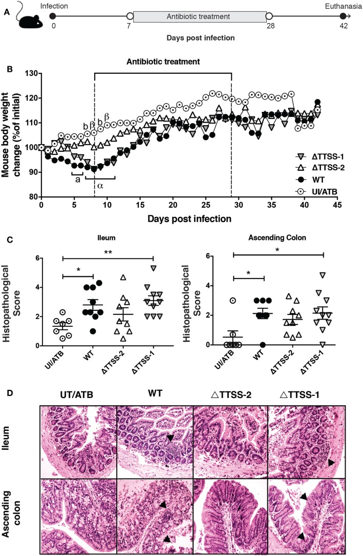
Figure 2.
Previous Salmonella enterica serovar Typhimurium (S. Typhimurium) infection increase the susceptibility of spontaneous intestinal inflammation of interleukin (IL)-10−/− mice. (A) IL-10−/− mice were orally infected with 1 × 10⁶ CFU of S. Typhimurium (STM) wild type (WT), ΔTTSS-1, or ΔTTSS-2. Seven days post-infection (days p.i.) mice were orally treated with enrofloxacin (2 mg/ml) for 3 weeks, left untreated for 14 days and euthanized to evaluate spontaneous intestinal inflammation. (B) Body weight were recorded on a daily basis for 42 days after infection. (C) Average histopathology score for ileum and ascending colon of 6–10 IL-10−/− mice included in each group. (D) Representative images of on the intestines of IL-10−/− mice infected with S. Typhimurium WT, ΔTTSS-1, or ΔTTSS-2 (4 µm ileum and ascending colon section stained with H&E and observed in optical microscope at 10× magnification). Body weight changes were analyzed using two-way ANOVA with Dunnetts post-test. Mice infected with WT (a, α) and TTSS-1 (b, β) were compared with mice uninfected and treated with antibiotics (UI/ATB). Data shown are mean ± SEM of three independent experiment with 8 mice uninfected; 10 mice infected with WT strain; 10 mice infected with ΔTTSS-1 strain; and 9 mice infected with ΔTTSS-2 strain. Statistical significance was indicated by letters: a, b: *P < 0.05; α, β: **P < 0.01. Histopathology score was analyzed by one-way ANOVA with Kruskal–Wallis post-test. Data shown are mean ± SEM *P < 0.05; compared with uninfected mice. Tissue damage and inflammatory infiltration are indicated (arrow).

