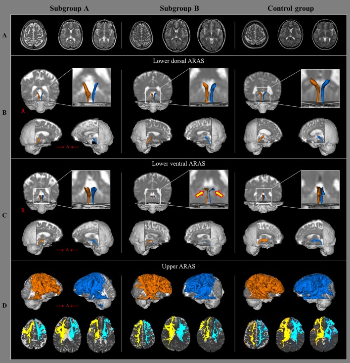
Figure 1.
Brain MR images and diffusion tensor tractography of the three components of ascending reticular activating system, in a representative patient and control subject. (A) T2-weighted brain MR images at the time of diffusion tensor imaging scanning in a patient with subgroup A (50-year old male) and subgroup B(35-year old male), and a control subject (63-year old female) show no abnormality. (B) Results of diffusion tensor tractography (DTT) for the lower dorsal ascending reticular activating system (ARAS). (C) Results of DTT for the lower ventral ARAS: the lower ventral ARAS in the patient with subgroup B is narrowed in the patient on both sides (arrows), compared with those of the control subject. (D) Results of DTT for the upper ARAS.

