
Fig. 2. Gene expression and kinase activity changes in BeWo cells under various stress conditions.
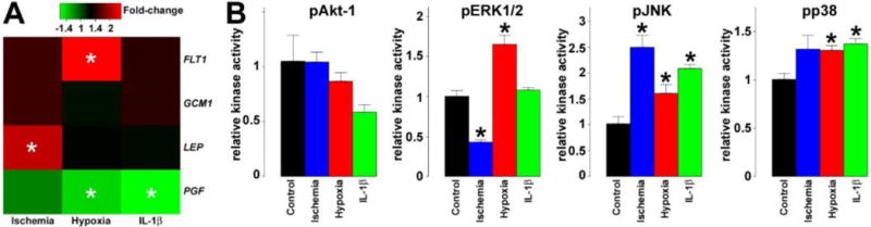
A) Heatmap representing qRT-PCR data reveals various effects of stress conditions on FLT1, GCM1, LEP and PGF expression in differentiating BeWo cells. The color bar depicts fold-changes relative to differentiating BeWo cells in normoxic conditions. In ischemia, LEP expression increased by 2.1-fold (p=0.04). In hypoxia, the expression of FLT1 increased by 2.5-fold (p=0.056), while PGF was down-regulated by 1.4-fold (p=0.04). After IL-1β treatment, PGF expression was decreased by 1.6-fold (p=0.01). B) Bar-charts representing kinase activity assay data reveal various effects of stress conditions on pAkt-1, pERK1/2, pJNK and pp38 activities in differentiating BeWo cells (n=3). In ischemia, JNK had a 2.5-fold increased activity (q=0.03), ERK1/2 had a 2.4-fold decreased activity (q=0.02), while there was a 1.3-fold, marginally significant increase in p38 activity (q=0.17). In hypoxia, the activity pERK1/2 (1.7-fold, q=0.03), pJNK (1.6-fold, q=0.1) and pp38 (1.3-fold, q=0.06) increased significantly. After IL-1β treatment, pJNK had a 2.1-fold increased activity (q=0.03), while pp38 activity was increased by 1.4-fold (q=0.04). All experiments were run in triplicate. Stars denote significant changes.