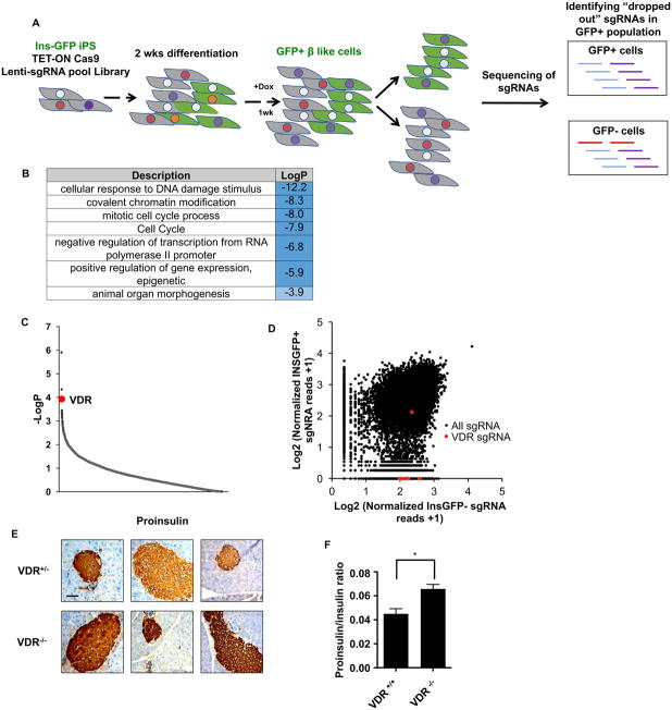
Figure 1. VDR is essential for β cell homeostasis.
(A) Schematic representation of the genome-wide CRISPR loss-of-function screen in human iPSC-derived β-like cells.
(B) Gene ontology analysis of targets compromising β-like cell function (p<0.01, 156 total genes).
(C) p-value rank order plot of genes enriched in the loss-of-function screen, VDR is labeled in red.
(D) Distribution of normalized reads of individual sgRNAs in GFP sorted cells; VDR sgRNAs are labeled in red. X-axis location indicates sgRNA were found only in INS-GFP− cells.
(E) Immunohistochemistry staining of pro-insulin in VDR+/− or VDR−/− islets (scale bar: 100μm, each panel represents an individual mouse).
(F) Serum proinsulin/insulin ratio in VDR+/+ and VDR−/− male mice measured by ELISA (n=3, mean±S.E.M, * p<0.05).

