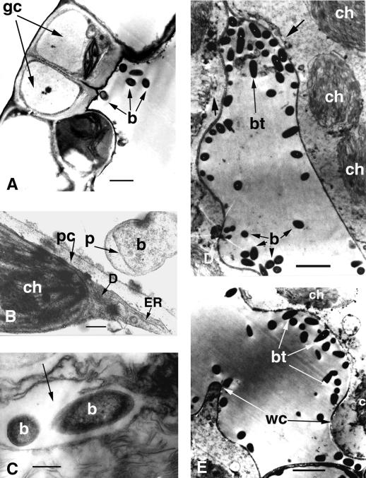
Figure 3.
Transmission electron microscopy of Arabidopsis Ll-0 leaves infected with P. aeruginosa PA14. Detached leaves were incubated in a bacterial suspension OD600 = 0.02 in 10 mm MgSO4 for 2 h under vacuum and then transferred to water agar plates. A, Cross section of a stoma and bacteria inside the substomatal cavity 24 hpi. Bar = 10 μm. B, Interaction of a PA14 cell with an Arabidopsis cell illustrating concentration of host organelles at the site of interaction with the bacteria at 24 hpi. Bar = 1 μm. C, Two bacteria shown inside host cytoplasm (arrow). Bar = 1 μm. D, Advanced stage of infection illustrating disrupted host membranes at 72 hpi. The bacterial thread has digested the middle lamella and separated the mesophyll cells from each other. The host plasmalemma (short large arrow) is highly undulated (left cell) or partly destroyed (right cell). The outer membranes of the chloroplasts are at various stages of degradation. Bar = 10 μm. E, Advanced stage of infection illustrating thinning of the cell wall and cell wall convolutions. The plasmalemma and chloroplasts are severely degraded. Bar = 10 μm. b, Bacterium; bt, bacterial thread; ch, chloroplast; D, dictyosomes; ER, endoplasmic reticulum; gc, guard cells; p, bacterial periplasmic space, pc, plant periplasmic space; wc, wall convolution.

