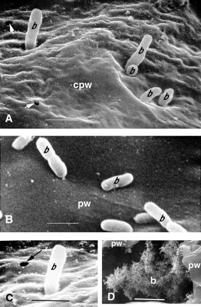
Figure 5.
Scanning electron micrographic visualization of freeze-fractured infected Arabidopsis Ll-0 leaves depicting P. aeruginosa PA14 cells attaching to and penetrating the convoluted cell walls of vessel parenchyma cells at 96 hpi. The leaves of intact Ll-0 plants were infiltrated with a bacterial suspension (OD600 = 0.02) under vacuum and detached leaves were incubated in Petri dishes on 1.5% (w/v) water agar as described in “Materials and Methods” for 72 h. A, PA14 cells on the surface of a highly convoluted cell wall of a parenchyma vessel cell. Holes in the plant cell wall with a diameter similar to that of the bacteria are apparent (arrowheads). Bar = 1 μm. B, P. syringae cells on the surface of a nonundulated cell wall of a parenchyma vessel cell. Bar = 1 μm. C, Detail of Figure 5A to highlight a hole in the plant cell wall. D, PA14 cells attached to the degrading cytoplasm in the lumen of a vessel parenchyma cell. Bar = 10 μm. b, Bacterium, cpw, convoluted plant wall; pw, plant wall.

