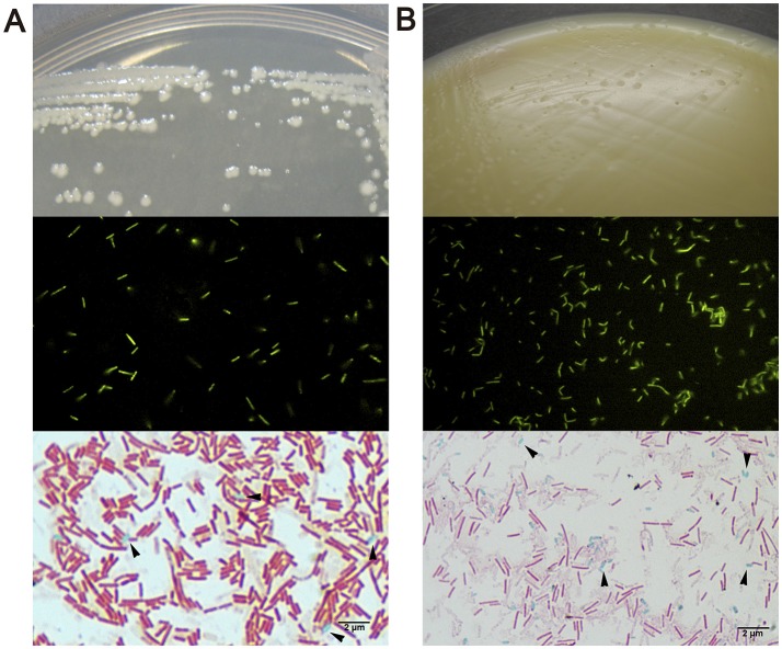
Figure 3.
Macroscopic and microscopic morphology of vegetative cells and endospores of (A) Bacillus clausii and (B) Clostridioides difficile. Upper show bacterial colonies in TSA or NN media. Middle show DTAF fluorescent bacteria. Lower show green malachite staining with black arrows indicating green endospores.

