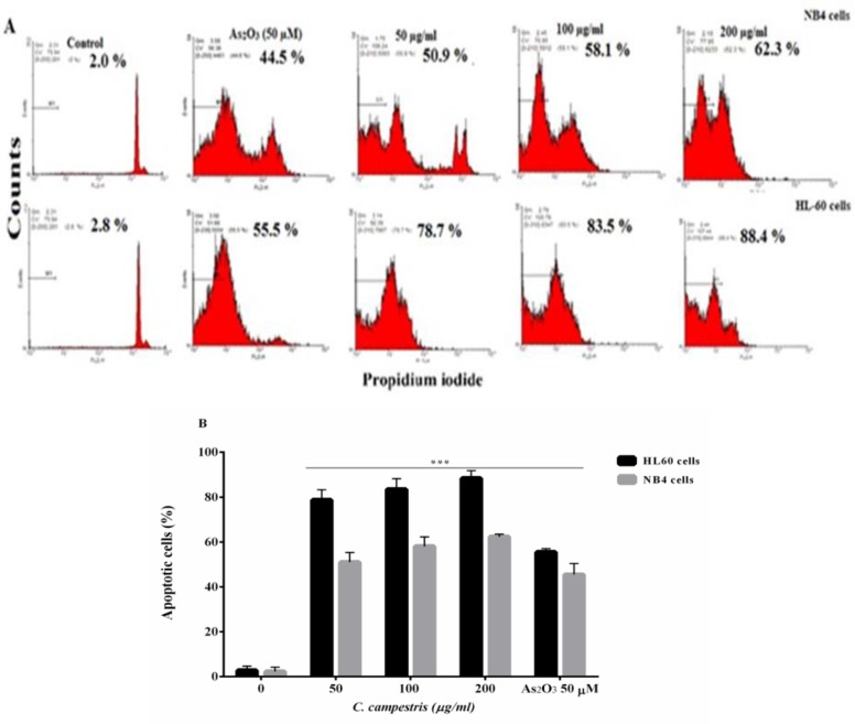
Figure 3.
Apoptotic cell death induced by C. campestris in leukemic cells. (A) NB4 and HL60 cells were incubated with different concentrations of C. campestris (12.5-200 µg/ml) or As2O3 (50 µM) for 48 hr. Apoptosis was assayed by PI staining and analyzed by flow cytometry. (B) Apoptosis rate shown as bars. The data shown are the means ± SEM of three independent experiments. ***p<0.001 as compared to control value

