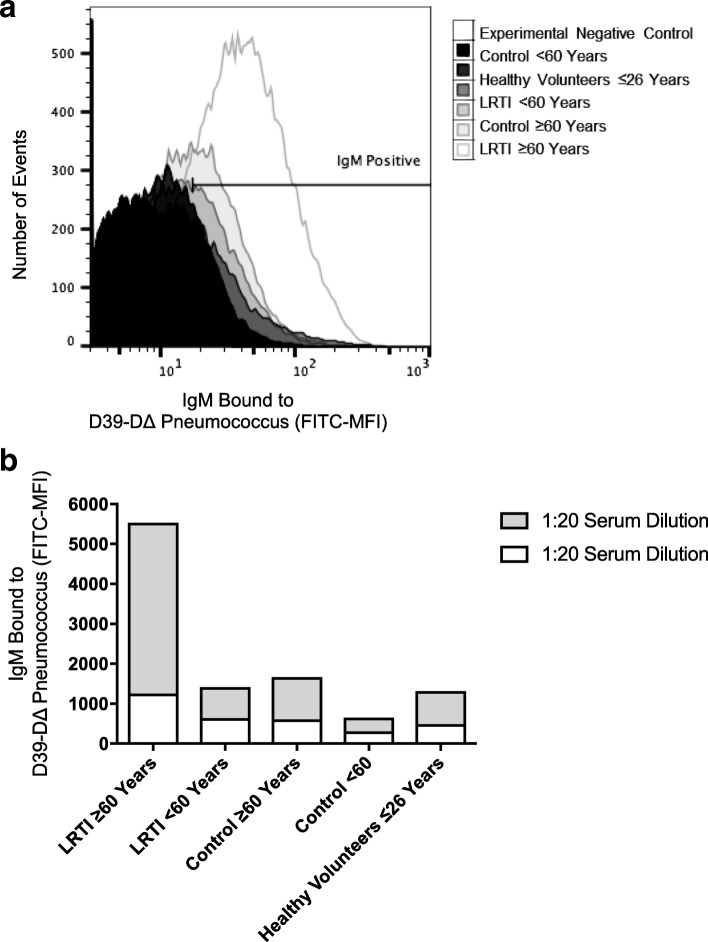
Fig. 3.
Flow cytometric assessment of IgM binding to pneumococcus surface. Pooled sera from different volunteer groups were added to bacteria. Two experiments using different dilutions (1:2 and 1:20) of sera were performed. a Histogram of surface binding of IgM to intact pneumococcus D39-D∆. b Surface binding of IgM from pooled serum samples expressed as the percentage of the bacterial population positive for IgM deposition multiplied by the geometric mean fluorescence of the bacterial population positive for IgM

