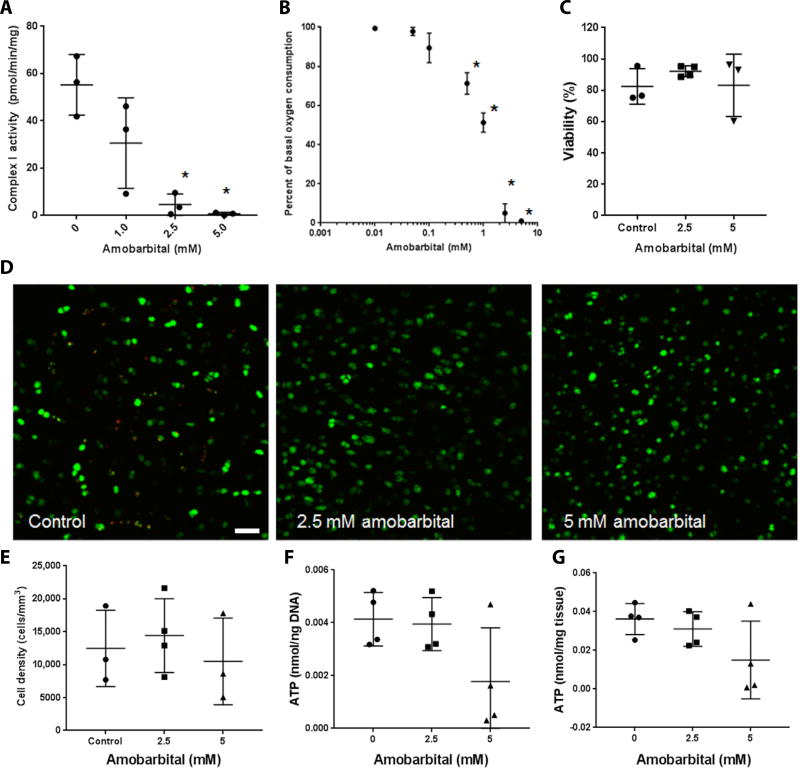
Fig. 1. Amobarbital inhibits chondrocyte complex I activity and is nontoxic to human chondrocytes at therapeutic doses.
(A) Spectrophotometric assay of freshly harvested bovine articular chondrocyte NADH (reduced form of nicotinamide adenine dinucleotide) dehydrogenase activity in the presence of increasing concentrations of amobarbital [n = 3; * P < 0.01 versus control via one way analysis of variance (ANOVA)]. For each group and panel, lines indicate mean and SD. (B) Extracellular flux analysis of oxygen consumption rates in bovine chondrocyte monolayers in the presence of increasing concentrations of amobarbital (n = 3; * P < 0.01 versus control via one way ANOVA). Circles represent means. (C) Percent viability as indicated by Calcein AM positivity of human articular chondrocytes taken from human intra-articular fracture (IAF) discards and cultured as osteochondral explants in the presence of amobarbital for 3 days (n = 3 except 2.5 mM, where n = 4; no significant differences via one way ANOVA). (D) Representative images of Calcein AM stained human cartilage explants exposed to amobarbital (n = 3; * P < 0.01 versus control via one way ANOVA). Scale bar, 50 µm. (E) Quantitation of viable cell density within cartilage from Calcein AM images of human explants (n = 3; no statistical differences indicated by one way ANOVA). (F) Whole human cartilage adenosine 5’-triphosphate (ATP) concentration normalized to total DNA content after amobarbital exposure (n = 4; no differences indicated by one way ANOVA). (G) Whole human cartilage ATP concentration normalized to protein content after amobarbital exposure (n = 4; no differences indicated by one way ANOVA).

